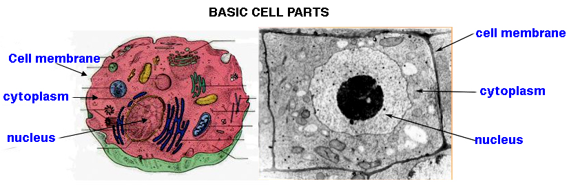
Cell
Organization: A cell has only
4 Basic Parts
-
-
1. cell
membrane -
selectively permeable - in/out.
(a
phospholipid bilayer)
2. a DNA
region
- (nucleoid or nucleus)
-
3. - protoplasm:
- a 19th century term for
vital fluids of cells
- cytoplasm:
- the
molecular skeleton giving form to a
cell
via cytoskeletal fibrous protein as
actins/tubulins,
and everything within a cell
- cytosol:
- is the
aqueous compartment without
organelles
-
4. -
organelles:
- 'mini organs'
within cells with specific
metabolic functions
-
*
-
-
So lets take a close look at the
structural anatomy of Cells...

-
-
-
-
-
-
There are only
2
successful Structural Plans of
Cell Organization
Prokaryotes
vs.
Eukaryotes
distinguished by
size & internal
structure &
presence/absence of organelles
Prokaryotes... Gk πρό- (pro-)
"before" + καρυόν (karyon)
"kernel = nucleus"
are
single-celled organisms without a
nucleus or major cell
organelles...
includes: bacteria,
archaea,
& cyanobacteria,
[bad puns &
jokes]
-
prokaryotes
are primitive, simple, yet
versatile, and a ubiquitous unicellular life
form
a
most successful evolutionary life
form-
making up about
2.5% of total biomass of planet Earth
over 2,500 different species
identified
(easily cultured
in lab)
500-1,000
species live in out human gut
guesstimates: 1% to 3% of
200 lb Human is bacterial
[i.e., about 2 to 6 lbs]
anim of
prokaryote structure &
characteristic
parts*
little to no
internal compartmentation (maybe a
few organelles, as
ribosomes)
lack complex
membrane bound organelles (may
have some specialized membranes*)
genome is "naked
DNA*" - i.e.,
no protein complexes & no real "chromosome?"
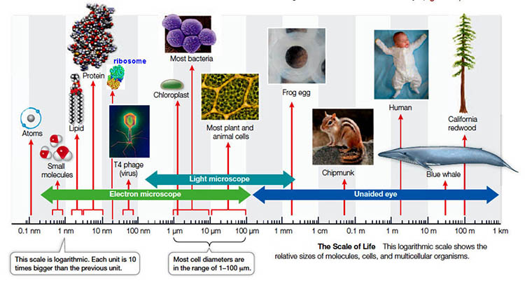
typical size range - 0.1 to 10 µm
diameter: Largest bacteria
discovered
--> size relationships*
&
Scale of
Life*
-
Procaryotes...
includes
all the UNICELLULAR
organisms of TWO
Domains :
the
Archaea
(Archaea
journal)
the
Bacteria
(Eubacteria
=
cynaobacteria,
mycoplasma, &
rickettsiae).
► Prokaryotes -
both archaea/bacteria evolved by
solving environmental challenges
and a versatile cell
chemistry, i.e., via novel &
new metabolic solutions...
1. ARCHAEA
- many living prokaryotic
archaea are called Extremophiles*
are organisms that
thrive in physical
or geochemical
extreme
conditions which seem
detrimental to the majority of
life on
Earth...
Extremophile
origanisms records*:
NASA found that after
almost 20 years of
continuous human presence,
the
International Space Station
has developed a core
microbiome of 55 different
microorganisms. As part of a
project called the Biology
and Mars Experiment,
researchers found that
bacteria, algae, lichens and
fungi survived on the
exterior
of the Space Station for 533
days.
...they
seem to be highly
conserved - "living
forms evolved for many
environments"
-
|
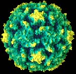
2. EUBACTERIA...
[ images
] "True
bacteria... modern day
microbes"-
includes
all living bacterial species and
cyanobacteria [excluding the archaea].
There are a
number of ways microbiologists
have categorized
Eubacteria:
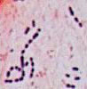
1- most exhibit 3 common
bacterial
shapes:
COCCI,
BACILLUS, SPIROCHETES
shapes
of bacteria*
(pics of bacteria
on pin & syringe
& staph aureus
infections)
2- many
microorganisms possess a flagella for
motility
fig of
flagella
3- can
also be distinguished by their cell wall
components via
Gram Staining*,
which can determine
what type of bacteria is present
& a treatment.
4- several eubacteria are
pathogens
and cause many human diseases*:
several Nobel Prizes have been awarded
for research on pathogenic-harmful
bacteria
yet, of the 2,500 species (? or more)
only 170 species are pathogenic in
mammals.
and many bacterial species also make
antibiotics, which kill other species
of bacteria.
History of
Antibiotics
& timeline of
antibiotics &
the Discovery of
Penicillin.
-
CYANOBACTERIA (are
eubacteria) - also called blue-green algae
(but no relationship)...
description - are
aquatic photosynthetic
unicellular Gram-negative
eubacteria (pics)
They have cytoplasmic
membranes & may catalyze N2
fixation [ N2 --> NH3, NO2-, NO3- ]
-
- EUKARYOTIC [Gk:
eu
-true karyon -nucleus...]
cell plan of MULTI-CELLULAR
ORGANISMS,
-
eukaryotes (eukarya)
include the fungi, algae, protozoa,
and all plants & animals all
contain
many
internal membrane bounded organelles...
organelle -
a subcell
part that has a distinct metabolic
function;
[akin to a
subcontractor on a construction job]
- Some common
CHARACTERISTIC of EUCARYOTES:
have a nucleus
- single greatest step in evolution
of higher organismal cells
genes
are in "chromosomes*" [colored bodies...
made of DNA + proteins]
contains more
DNA (1,000x
more) than prokaryotes: linear vs.
circular.
presence of less
cell "wall"
structure with a more flexible extra-cellular
matrix
extensive internal
membrane systems
elaborate cytoskeleton - provides
internal framework; favors larger
cells
presence of organelles -
significant internal
compartmentalization of functions
reproduce
sexually
usually larger -
cell volume
10X > than bacteria - size 5.0 to
20 µm diameter
2 major kinds of
eukaryotic cells are commonly
recognized:
-
-
animal -
metazoan
cell*
- heterotrophic
metabolic feeder
-
plant -
metaphyta
cell*
- autotrophic
producer
- contain
chloroplasts, large vacuoles, &
a cellulosic cell wall
-
-
Procaryotes vs. Eucaryotes
-
-
table of similarities &
differences*(later)
How do research
biologists study & identify the
subcell parts, - the Organelles
|
|
- 2. Key
Functions of Microscopes:
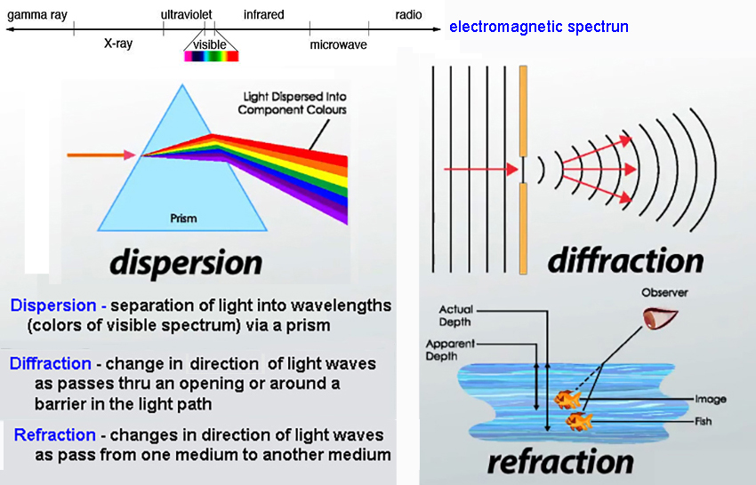
Optical microscopy involves the diffraction, refraction, or dispersion* of light (electromagnetic
radiation) interacting with live or prepared
samples &
subsequent collection of scattered
radiation
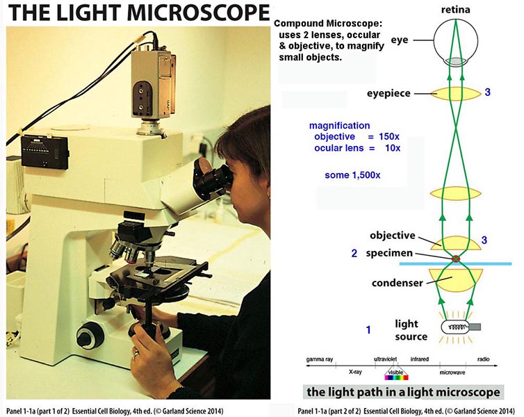
(light) to build up magnified images of small objects using optical compound lenses*
Magnification =
objects appear larger in a light microscope =
typically about 1,000 fold
Resolution*
= minimum distance between objects
that can still be seen as 2 dots...
resolution of human
eye
= 0.2 mm (200um)
resolution of light
microscope
=
0.2
µm (200nm) (1,000x human eye)
[ resolvable size
scale & scale of biological parts* ]
Killing/Fixing of
samples*
: specimens are preserved
from decay and autolysis;
ongoing biochemical reactions are stopped, &
proteins denatured & 'fixed'
in place;
formaldehyde & glutaraldehyde
denature all of a cell's proteins
increasing mechanical stability, but may
produce artifacts* in
microscopy.
Embedding
& sectioning : Cell water is
replaced by a more rigid paraffin or plastic
material
and sectioned by a microtome* (1 to 10 µm thick tissue
sections)
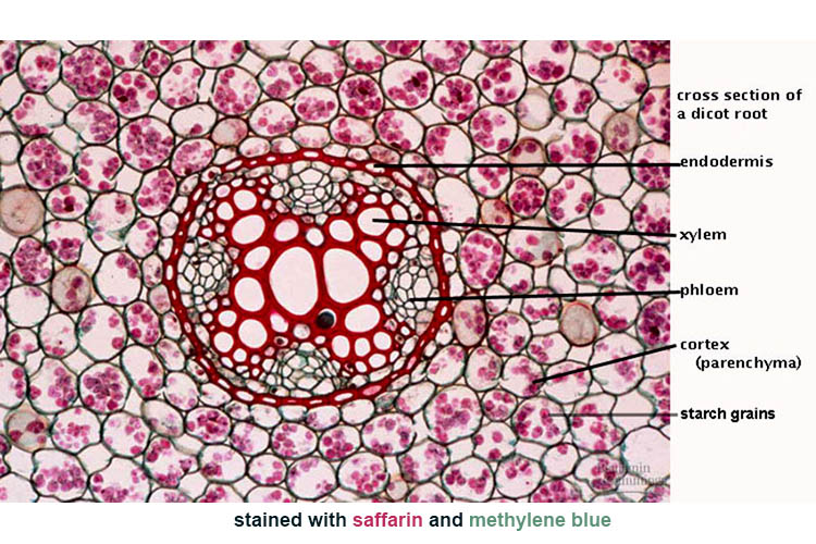
Selective
staining:
stains (dyes) attach to
specific molecules colorizing them (root*)
Types of
Light Microscopy*
2. Electron
Microscopy...
How electron
microscopy allowed identification of subcell
parts...
animation on how EM
works*
and
a primer on Electron Microscopy
George
Palade (NP-1974) - pioneered use of EM for
studies of cell structure
resolution* =
0.2 - 2.0 nm -
(scale of biological parts & specimen preparation*)
image analysis... pic
of TEM scope 3D
restructuring*images*
an analogy = Mark Light or Hard Rock
4 types of Electron
Microscopy include:
TEM -
Transmission: a TEM micrograph* (often stained w
osmium tetroxide
binds to lipid membranes)
SEM -
Scanning: image from 2ndary e's emitted from electron beam
on a metal shadow cast
comparison* - sample
is killed/fixed/dried - coated
with gold/palladium* for output
of 2ndary e's.
the typical resolving power is about 2 nm (1/10 of a
ribosome)
some examples* &
lily pollen grain & a Tardigrada
winner
FFEM - in Freeze Fracture
EM samples are frozen & cracked along plane
of least resistance,
usually along
hydrophobic membranes. how to prepare*, results*, & TEM v. FFEM*
Cryo-EM
- protein
samples are cooled to cryogenic temps (-2000C)
in vitreous water to observe
via
narrow e- TEM beams with multiple
images of each electron to reconstructe
its biomolecular structure at near atomic
resolution (2nm). [protocol*
& 3D-fig*].
- Model
Biological Experimental Systems for use with
Light & Electron Microscopy include...
a.
isolation and/or
culture of cells: RBC
cells & 1st Human Cells cultured-HeLa cells*
b. Cell Fractionation
& differential Centrifugation*
is
used to isolate cell organelles
c.
3D cell imaging*view@home
---> Allen
Cell Explorer
-
Summary examples of
microscopy images*...
Microscopy has
given us views of the major eukaryotic
organelles
[common ex:
epithelial cells]
NUCLEUS
:
membrane, pores, chromatin, nucleolus, nucleoplasm
MITOCHONDRIA
:
peri-mitochondrial space, cristae, mitoplasm
(matrix)
CHLOROPLAST
:
peri-chloroplast space, thylakoids, chloroplasm
(stroma)
RIBOSOME
:
small subunit, large subunit, polysome
ENDOPLASMIC
RETICULUM : smooth
& rough
GOLGI BODY
:
cis & trans sided - endomembrane pathway
LYSOSOME
:
hydrolytic enzymes
MICROBODIES
:
peroxisome & glyoxysome
CYTOSKELETON
:
microfilaments, microtubules, intermediate
filaments
CENTROSOME
:
centriole, basal body, flagella, cilia
INTERCELLULAR
JUNCTIONS :
tight junctions, desmosomes, gap junctions,
plasmodesma
PLANT CELL
VACUOLE
:
tonoplast, cellular waste, and osmoregulation
CELL
MEMBRANE
:
selective transport barrier.
Gram Staining is
a method for the differential staining of
bacteria:
samples are stained in a solution of crystal
violet,
& then treated with
iodine
solution, rinsed, and then
counter-stained with safranin
O;
The staining differences are due to
the bacterial cells wall structure
Gram staining is due to a
differences in cell
wall structure*
&
is useful
in identification & determining an
appropriate treatment for an infection.
back
|


