Research Methodologies of CMB - from 1920 to 2020
Instrument advances within this 100 year
period are the epitome of modern scientific
age.
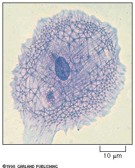
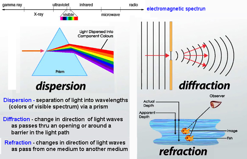
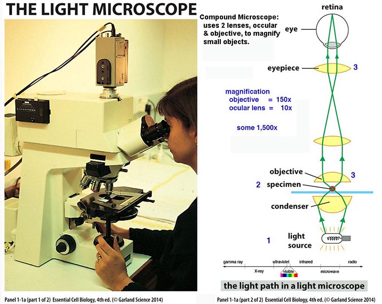
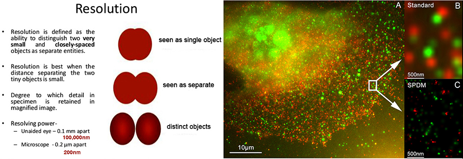
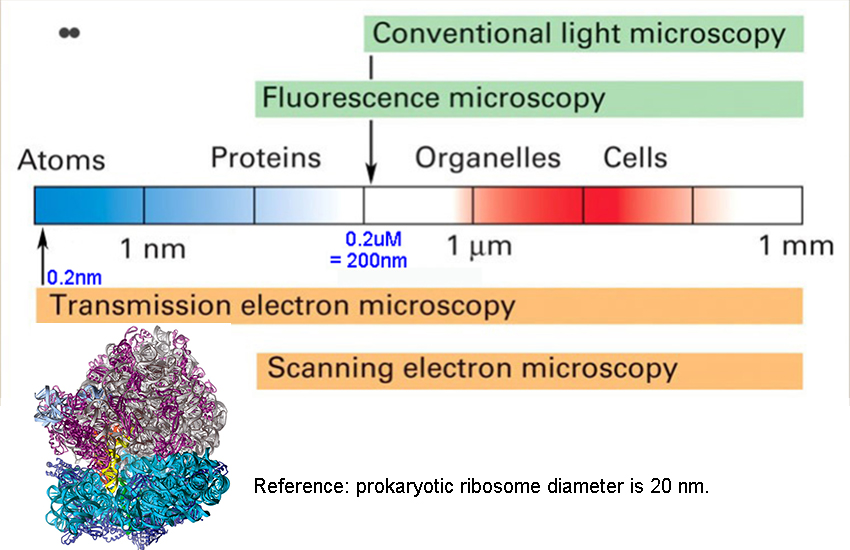
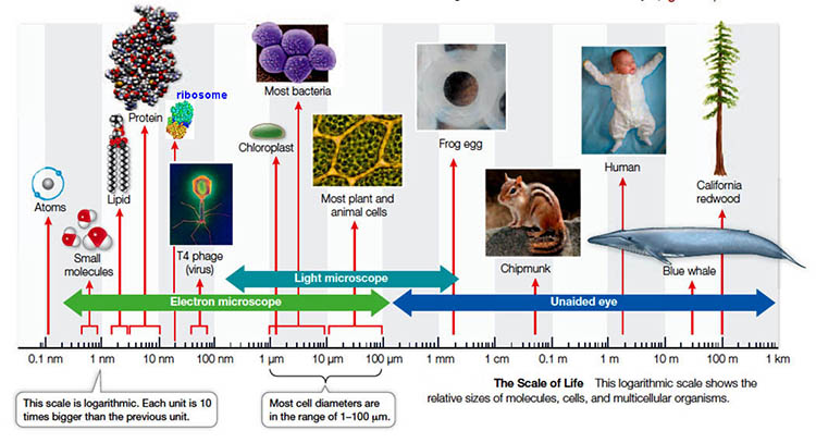
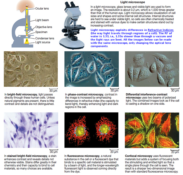
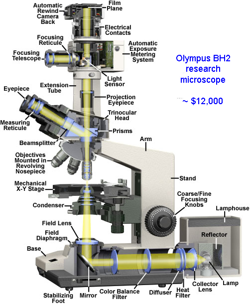
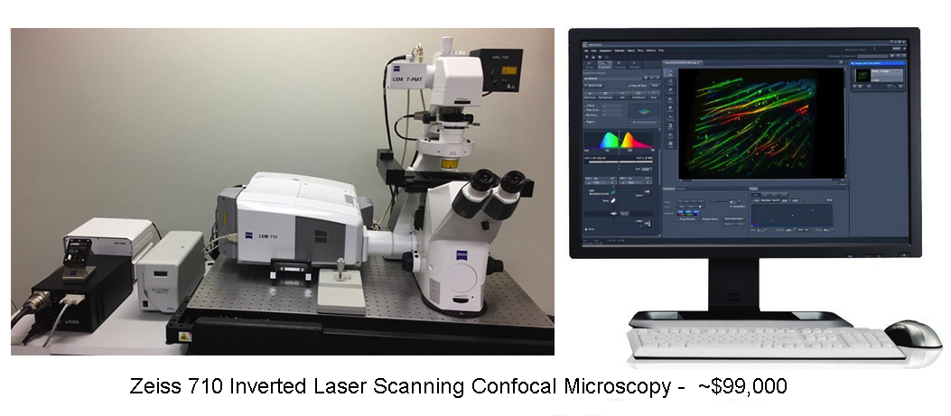
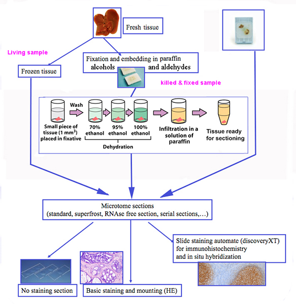
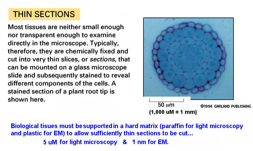
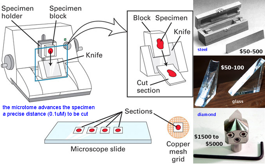
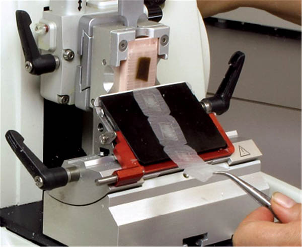
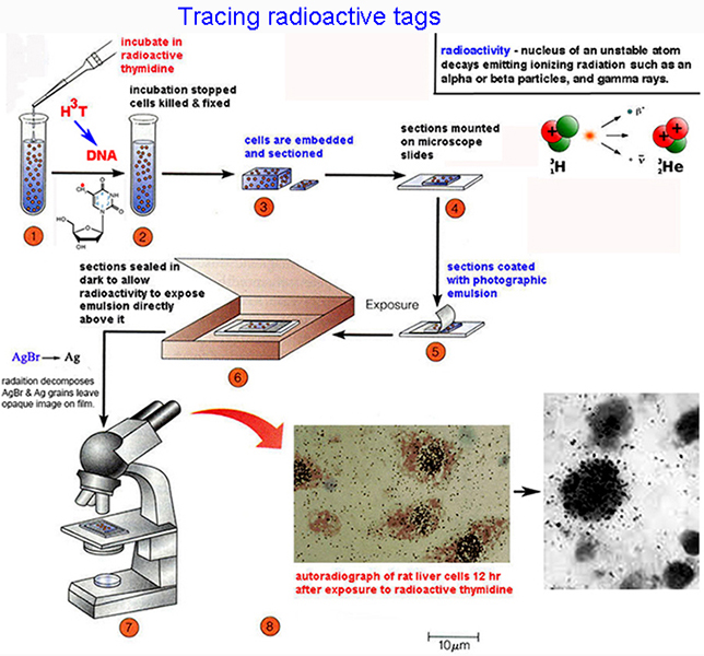
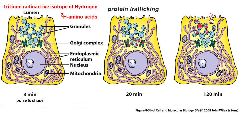
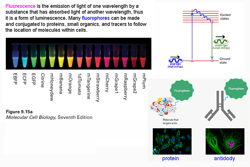
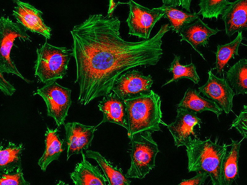
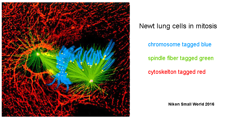
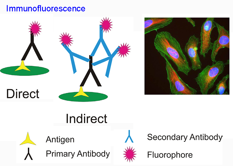
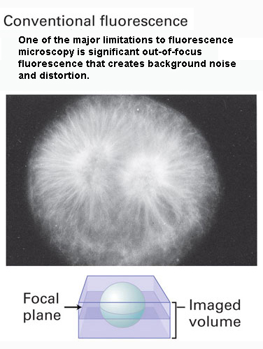
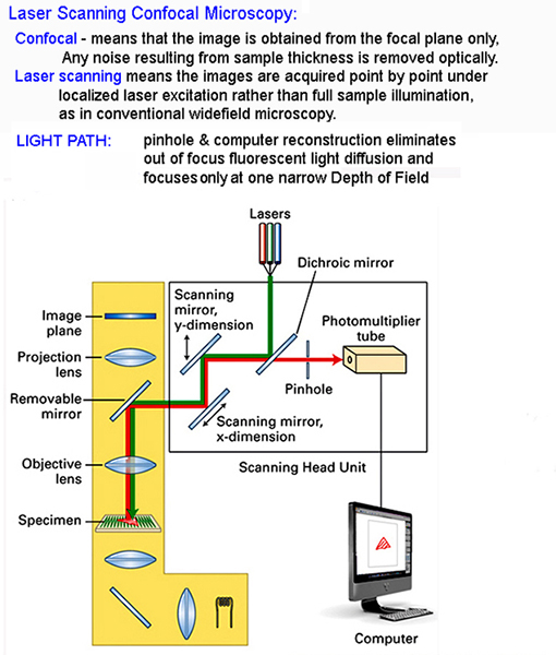
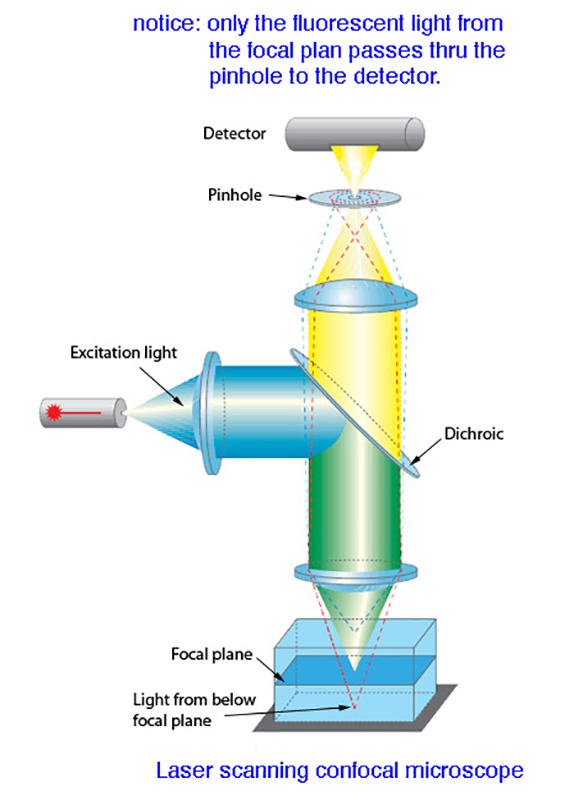
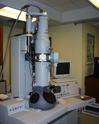
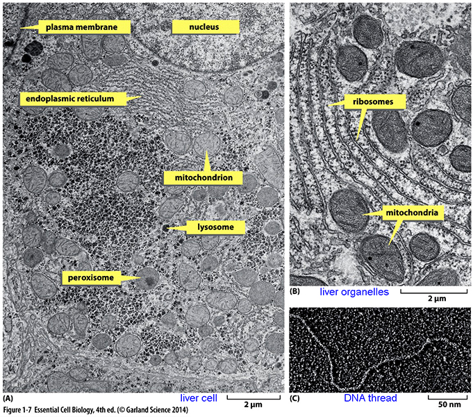
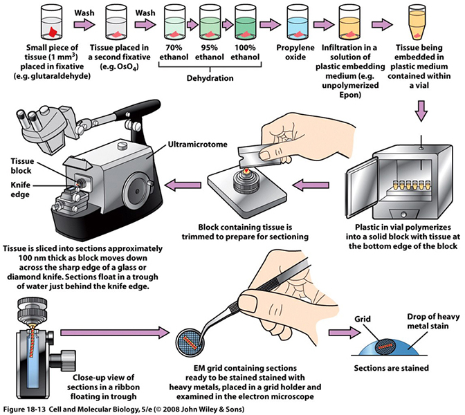
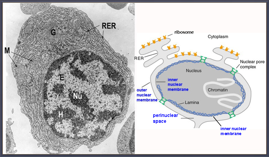
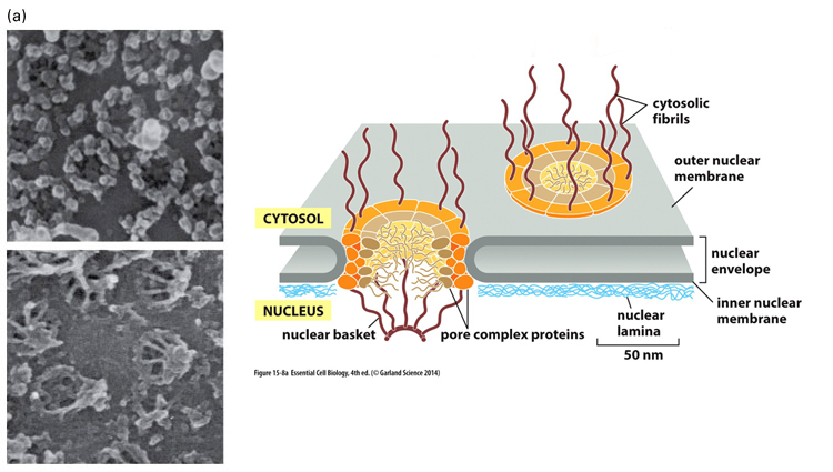
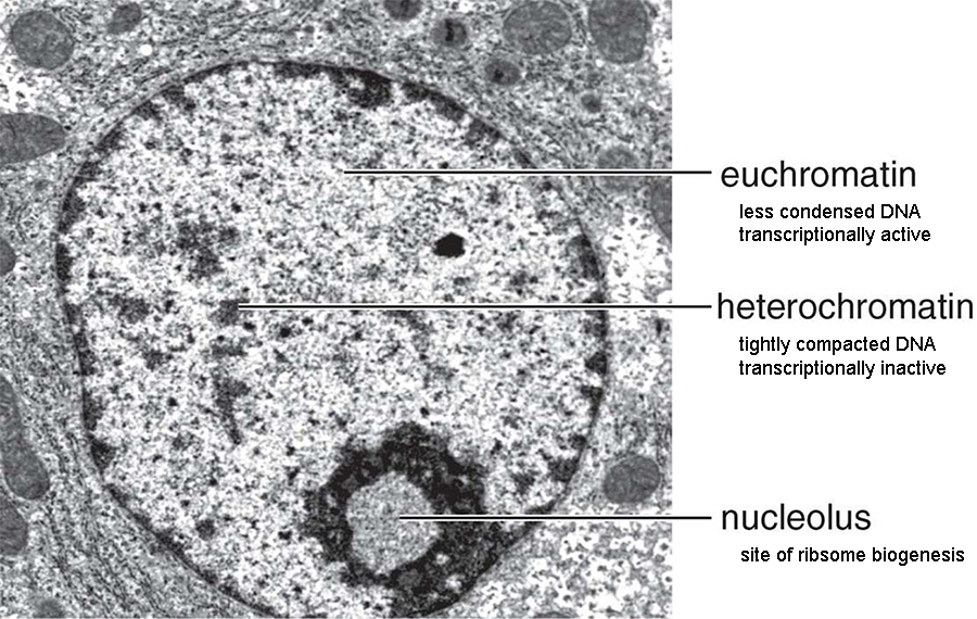
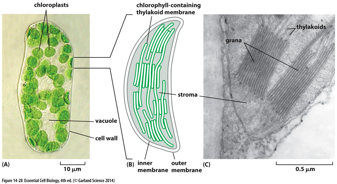
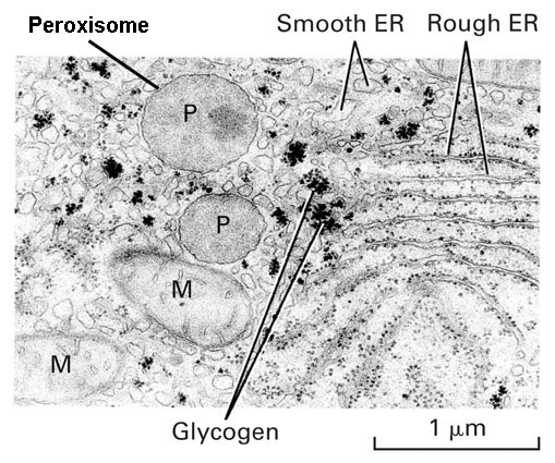
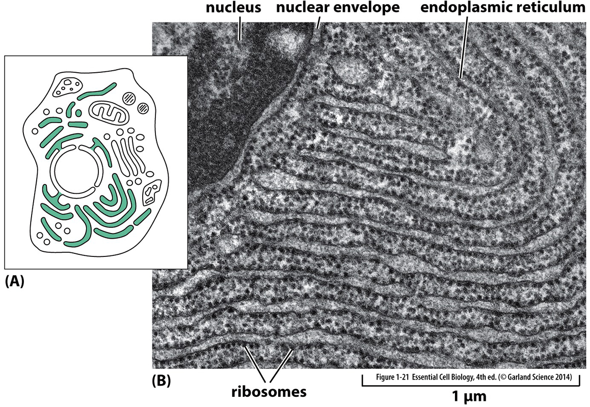
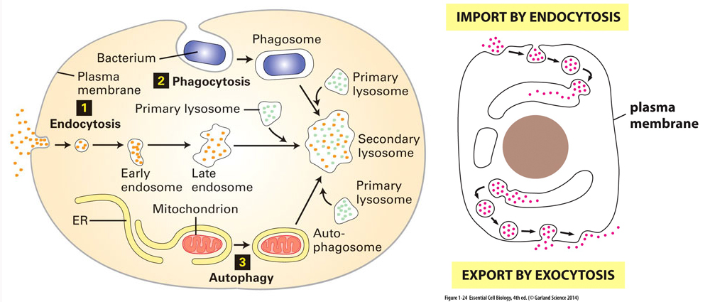
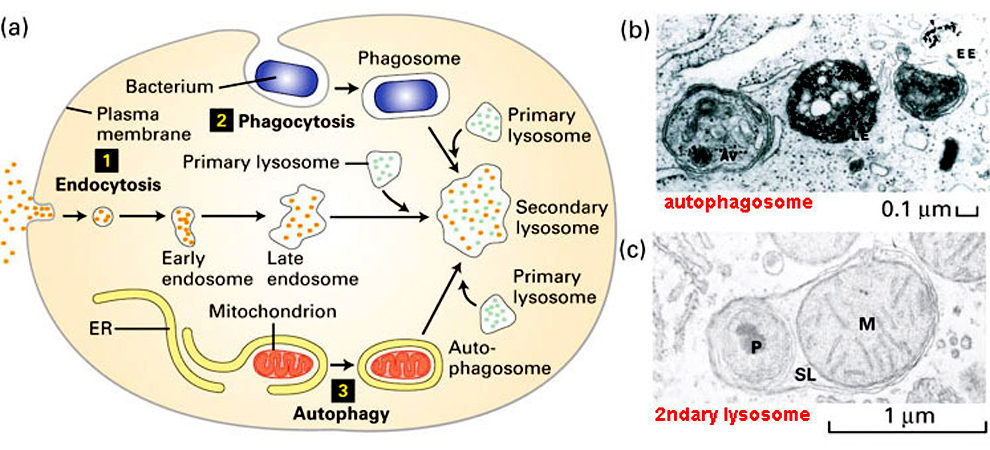
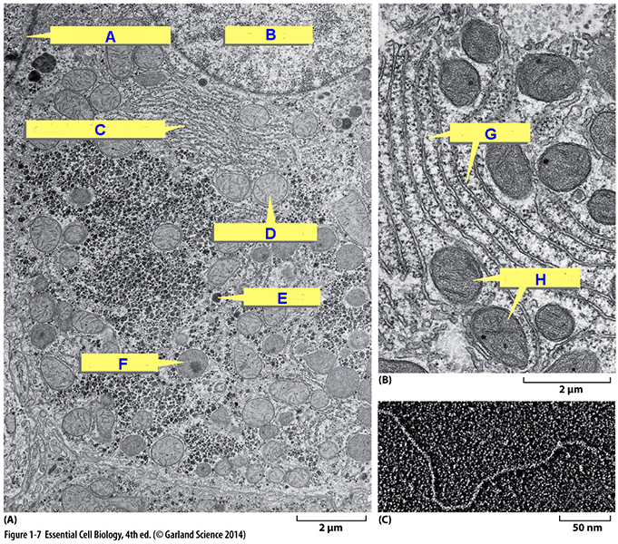
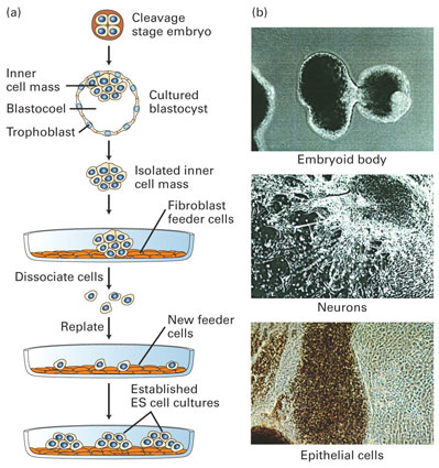
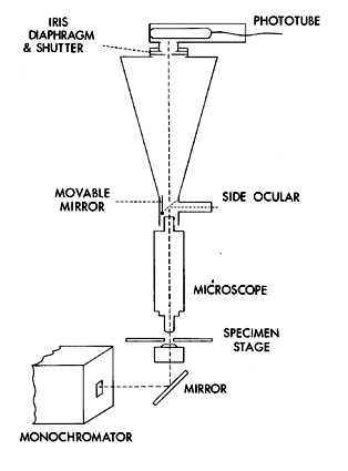
MICROSCOPY is the optical discipline using microscopes to view small cellular objects: the development of microscopy revolutionized biology and remains an essential tool of CMB. 2 Types of Microscopy: Light (optical) Microscopy & Electron Microscopy Light Microscopes* - optical microscopy involves the diffraction, refraction, or dispersion* of light (electromagnetic radiation) interacting with live or prepared samples & subsequent collection of scattered radiation (light) to build up magnified images of small objects using compound lenses* i.e., an objective lens - magnifies object (150x) & ocular lens (10x) = 1,500x magnification. 1857 Zeiss sells 1st compound scope 1872 Abbe partners with Zeiss & optimizes microscope designs (lens & condensors) - lens resolution* is 0.2 μm or 200nm*: near limits of light - Microscopy-Airy disks - Scale of Life* - relative sizes of molecules, cells, and multicellular organisms. ► types of light microscopy* --> (for more info on the basic concepts - microscopy resources) ► student microscopes - AmScope vs. research microscopes - Olympus BH2 & Zeiss 710 ► specimen preparation - living vs. fixed materials. killing, fixing & embedding* --> physical distortion via fixatives* Tissue Sections must be very thin 1-50um - microtome* & sectioning* $-$$ Some Light Microscopy Methodologies... ecb panel 1.1 pg 10-11 ► tagged precursors can trace molecules in cells & biochemical pathways: Autoradiography - 1940 S. Warren produced an autoradiograph, which is a microscope image on film (in emulsions of AgBr) produced by a pattern of radiation decay (e.g., beta particles or gamma rays) that can reveal the distribution of location of the labeled material in a cell. methodology & preparation*... one can also track proteins* Fluorescence microscopy* [2008 Nobel] a form of light microscopy where a cell part has been specifically labeled with a fluorescent molecules* (1st tag was Green fluorescent Protein). ► examples of fluorescent images = fibroblast & mitosis & Limbs immunofluorescence microscopy - uses antibodies fluorescently tagged to bind specifically to a corresponding antigen as a probe for identifying a particular molecule in cells, tissues, or tissues, or biological fluids: ex. rat intestine* and focal plane problems* confocal fluorescence microscopy*- 1957 Minsky - confocal microscopes uses pinpoint illumination of fluorophore in one focal plane to eliminate out-of-focus fluorescence. A computer reconstructs the image and since only fluorescence in a narrow focal plane is detected, image resolution* is greatly enhanced providing sharper image... $ Recent advances in Light Microscopy & Olympus BioScapes microscopy Imaging 1998 cell imaging station - fluorescent-labeled sample detection and cost. 2015 expansion microscopy - allow resolution to 70 nm levels vs. normal 200 nm. ELECTRON MICROSCOPY - uses a beam of electrons to create an image ecb panel 1.1 page 11 wavelength of electron beam is 100K shorter than light rays = resolution in 0.2nm range TEM - Transmission Electron Microscopy - (electron optics): 1933 Ruska (1986 Nobel) 1st Transmission Electron Microscope TEM-principles* & ecb4e 1.7a* TEM passes e's through a specimen onto a viewing screen - Animation of how an EM worksEM*
(magnifies million fold,
theoretical resolution = 0.005
nm, but effective resolution
is = 0.1
to 0.2nm or
larger).
1952 Porter
/ Palade
- 1st TEM pics & EM stains - imaged
via scattering of e's by molecules
within
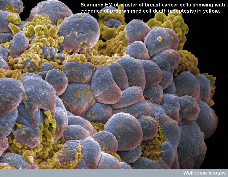
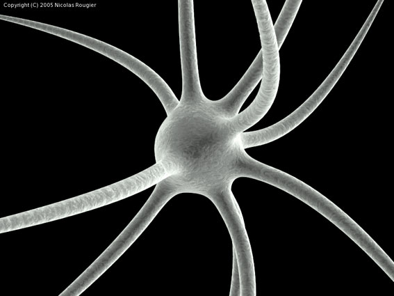
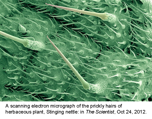
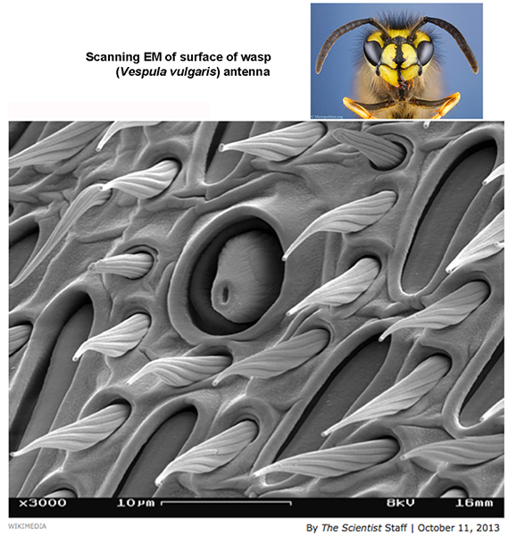
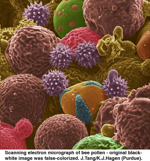
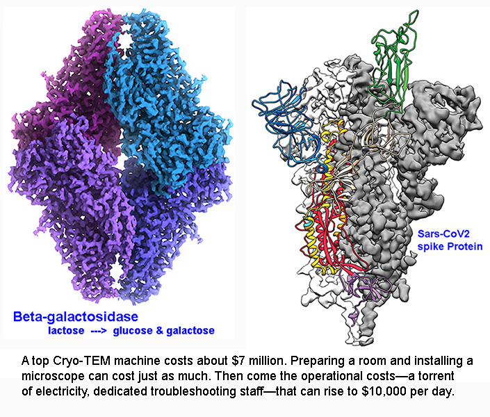
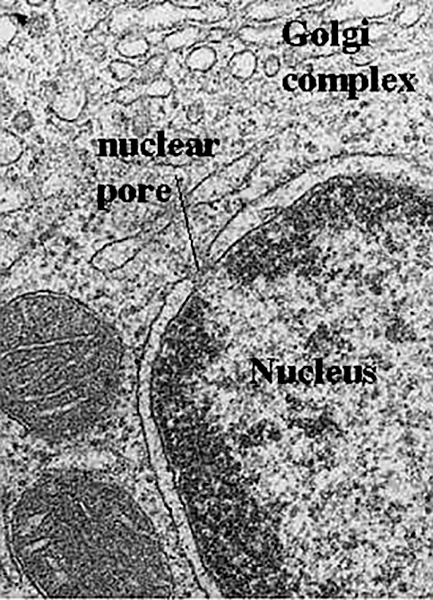
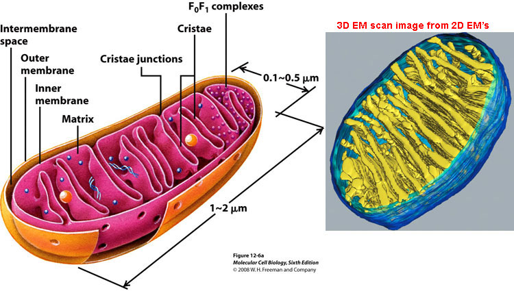
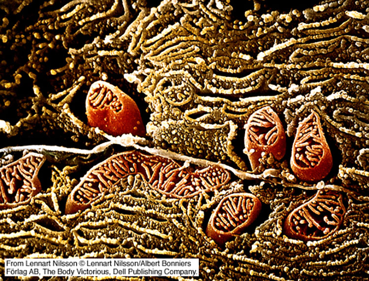
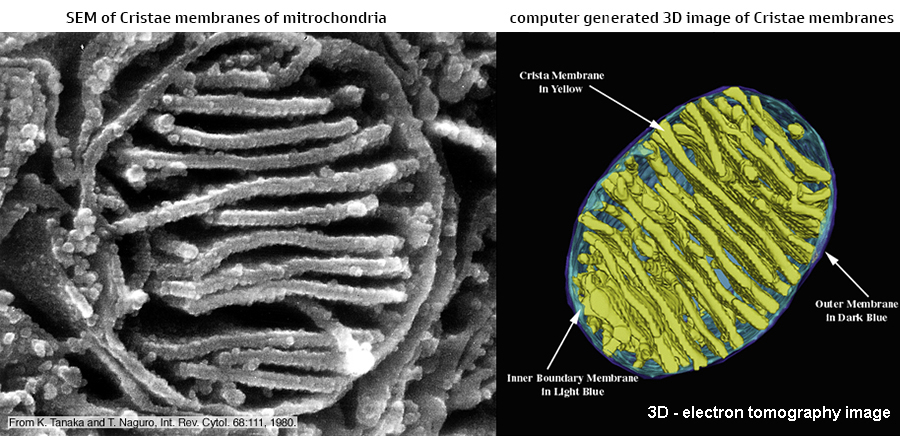
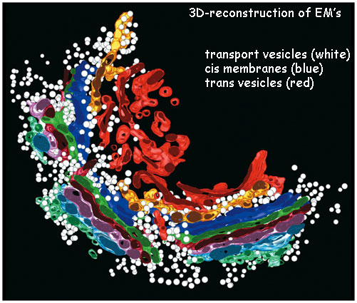
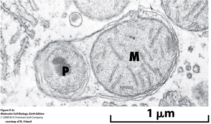
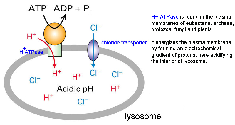
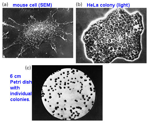
specimen (heavy metal stains as osmium tetroxide for lipid membranes) stain = dark. specimens must be very thin = 50 nm or less; sections are cut via microtome. 1957 Robertson - unit membrane hypothesis (all membranes look alike* in EM) 1981 Tagging - antibody tagging with gold particle in electron microscopy - fig 9.21* 2001 computer image averaging allows 3D modeling: Tomography* = ribosome & Ca-ATPase pump* fFreeze Fracture EM - : samples are frozen, metal coated, & cracked along plane of least resistance, usually along hydrophobic membranes. 1964 Steere & Muhlethaler - develops freeze fracture EM - prep* & pic1* SEM - scanning EM: : provides a great depth of focus with detailed images of cell surfaces & organisms. 1965 Charles Oatley - 1st scanning EMs are published. uses metal shadowing & coating* of specimens to reveal surface topography images* specimen bombardment releases 2ndary electrons - ecb panel 1.1* when focused onto detector reveals 3D surface details... mcb fig 9.25* & cancer cells some examples - neuron & plant & antenna & pollen & Hitachi Table Top model & Eye of Science Cryoelectron Microscopy - [NP 2017] - aqueous specimen is frozen in liquid nitrogen (-2000C) protocol* specimens retain native shape (no fixatives) allowing minute structural details* ThermoFisher Tundra Cryo-TEM to image large, biologically complex structures [cost].
The light
microscope, so called because it employs
visible light
to detect small objects,
The electron microscopy uses a focused
electron beam on fixed sectioned of cells,
which
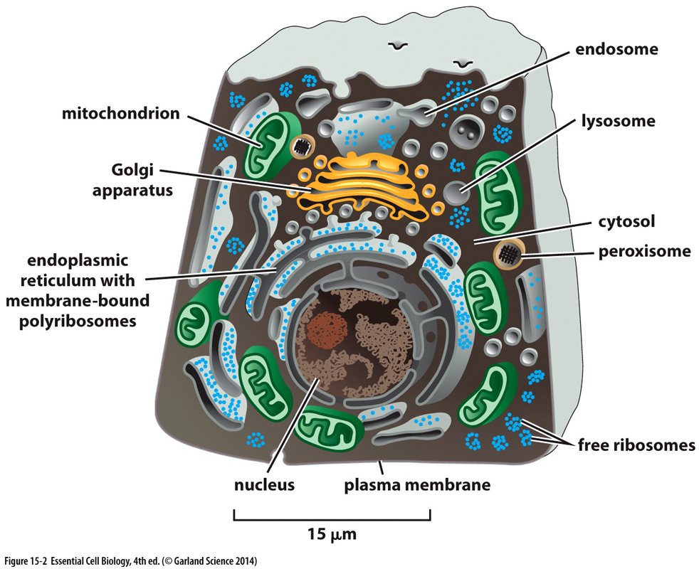
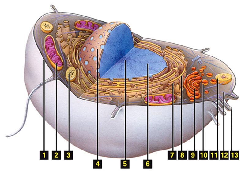
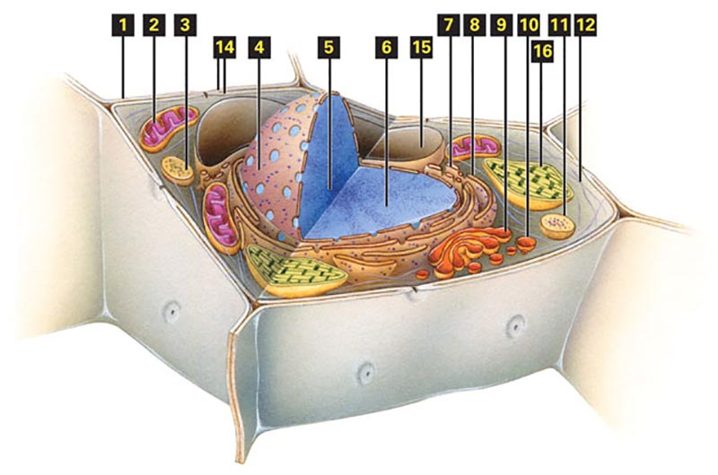
The material below on CELL ORGANELLES* is a
general review of your
4. endoplasmic
reticulum... network of
closed-flattened membrane sacks called
cisternae
"The
Inner Life of a Cell " - a movie animation by David Liebler
& Harvard U.
Download Power
Point
Please skip all the material below this point...
Where does one
get homogenous cell populations foe
microscopy???
http://micro.magnet.fsu.edu/primer/java/electronmicroscopy/magnify1/ How to video on electron cryotomograph
Modern
Cell Biology
is often dated from the works of
G.
Palade,
C.
deDuve,
A. Claude
|
|
|||||||||||||||||||||||


