MUSCLE
PHYSIOLOGY...
a
correlation of Form vs. Function
The response of a Reflex Arc
& Correlation of Structure & Function...
Campbell 12e reads: C50: (pg
1125-1132) & Campbell 11e
reads: C50 (pg
1123-1129).
Model: vertebrate skeletal neuromuscular
junction
- striated
skeletal muscle cell*
pic
myoblasts (muscle cells)
- innervated
muscle fiber* pic
neuromuscular junction
Muscles
cannot stretch (pull),
they may only
CONTRACT
(get shorter via a pull)
contractions provide forces that move
bones acting as levers (fulcrum point).
A
muscle CONTRACTION is
also called a muscle TWITCH...
| 4 parts of a Slow Twitch
Muscle Contraction CONTRACTION CYCLE* |
| 1) latent period
-
5 msec |
time between
application of AP
& initiation of contraction |
| 2) contraction
-
40 msec |
muscle shortens &
does its work |
| 3) relaxation
-
50 msec |
muscle elongates &
returns to original position |
| 4) refractory period
- 2 msec |
time of recovery between
stimulations of muscle |

some common Properties
of muscle contractions (twitches)...
Summation -
a 2nd contraction before 1st subsides tetany* (cause:
time differential of nerve/muscle)
Fused
tetany -
contraction of muscle remains constant without
relaxation
Fatigue - under repeat stimulation,
contractions get feebler, lactate accumulates,
pH changes lead to stopping of contractions
Charley Horse -
severe muscle cramps caused by pH imbalances, low Ca
levels, & dehydration.
Shivers
- involuntary-summed muscle contractions, which
releases waste heat, that warms body
Major
Muscle Types... ability of muscle to contract is
based upon MUSCLE PROTEINS*...
44 major types
of muscle proteins: 1. MYOSINs,
2. ACTINs,
3. Troponins,
& 4. Tropomyosins...
1.
Myosins are often Type Classed based upon the
MYOSIN
family protein
fibers present:
(also called heavy meromyosins*):
MYOSINS
are motor*proteins*
that move on actin filaments, whereas kinesin and
dynein
motors move on microtubules. Upon
interaction with actin filaments, myosin II uses
energy from ATP
hydrolysis to
generate a mechanical force for movements.
Myosin
isoform types are conserved
evolutionarily:
Comparing myosin
isoform types from different mammals
reveals remarkably little variation within a type
from
species to species, i.e., Rat Type I is more similar
to Human Type I
myosin, than it is to Rat
Type II's.
Thus selective evolution has maintained a functional
difference between Type I's
& Type II's over
eons of evolution.
Besides classifying
muscle based on the type myosins
present (different
isoforms)
muscle are often classified on the speed of
contractions into 2 Muscle Fiber Types:
which are
determined both genetically &
functionally.
TYPE I
(Slow Twitch
muscles)
&
Type
IIa/IIx (Fast Twitch
muscles)
Classification is based
upon how fast they can produce a
contractile twitch.
all
muscles are composed of varying %
composition of these two types |
| SLOW
TWITCH - TYPE I |
FAST
TWITCH - TYPE
II (IIa &
IIx) |
| slower
contraction times (100-110 mSec) |
faster contraction times (50 mSec) |
| tonic
muscles (darker:
red) - leg muscles |
tetanic muscles (paler: white) Pectoral (chest) muscles |
| contain myoglobin (red) |
no myoglobin (white) |
continuous use muscles - prolonged
performance
...endurance performances ( marathoners) |
one time use muscles - brief performances
...
for power & speed (sprinters) |
* predominantly AEROBIC enzymes
& metabolism
... cell respiration pathways
|
* predominantly ANAEROBIC (glycolysis)
... easily converts glycogen to
lactate w/o O2 |
| marathoner: 80% type I
& 20% type II |
sprinter:
20% type I & 80% type II
|
Distribution of Slow
& Fast Twitch muscle in Humans
|
| tropinin has lower affinity for Ca
|
troponin - higher affinity for Ca |
Relative
Distributions of Slow Twitch & Fact Twitch
Skeletal muscle Subtypes - (Type I & Type II) |
| |
Type I
(slow) |
Type II
(fast) |
Type IIa |
Type IIx |
| Average person |
50% |
50% |
40% |
10% |
| sprinter |
20% |
80% |
45% |
35% |
| marathoner |
80% |
20% |
20% |
0% |
| couch potato |
40% |
60% |
30% |
30% |
| spinal injury |
4% |
96% |
48% |
48% |
Due to
the conserved
evolutionarily stabilty of myosin
types they can not be converted,
thus a spinal cord
injury cannot be repaired by converting Type
II myosins to Type I's.
back |
Molecular Basis for Muscle
Contraction & Structural Model of Muscles
Vertebrate
Skeletal Muscle cells are multinucleate cell*
- muscle diagram
|
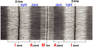
the
SARCOMERE is a basic repeating structural unit
of striated muscle...
and is defined by its banding appearance
in microscopy: delimited by Z-lines...
I band
- "paler zone" around Z-line
(Isotropic - passes light in all
directions)
A band
- "dark region" in center of sarcomere (Anisotropic - in
different directions)
M line
- "denser" mid point of the sarcomere
H zone
- "paler zone" in the center of sarcomere
around M line
|
 myofibril*** myofibril***
|
SLIDING FILAMENT
THEORY of Muscle Contraction (Hugh Huxley-1954)
I band varies
in length becoming shorter &
disappearing during contraction
A band remains constant in
its
size dimensions
contraction**
H Zone becomes denser
during a muscle contraction |
|
relaxed/contracted* & at
the molecule level - actin/myosin* contraction
animation* |
simple animation &
animation*
|
The 4
Major Muscle Proteins interactions are
responsible Contractions
|
|
|
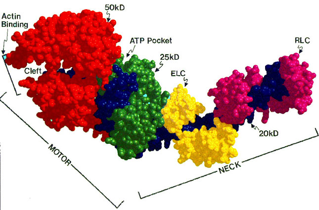
1. THICK
FILAMENT (A band - dark)
myosin II
- a dimer twisted to form 2 helical fibers with
globular heads,
each of which has
ATPase activity*
& an affinity to bind to actin
|
2. THIN FILAMENT
(I band - light)
F-actin* -
globular protein which polymerizes
into polymeric helical fibers...
each globular actin unit
contains a myosin binding site |
3. Tropomyosin* - fiber-like
protein which wraps
helically around thin filament
covering
the myosin binding sites on actins |
4. Troponin* - globular
protein complex which binds Ca+2 & initiates
contraction cycle
is a complex of 3 proteins,
Troponins
C,
I, &
T, which
bind Ca;
Troponin C (18 kD) binds Ca
reversibly...
Then TnC
binds TnI (23
kD) & TnT (37
kD),
which change their conformations in response
to TC binding Ca
causing tropomyosin to open the myosin binding sites
on actin. |
| |
https://www.youtube.com/watch?v=Ut-6pyIN7QE
SKIP All the MATERIAL from this
point below
Anabolic
Steroids & muscle physiology
& Doping and Muscle
Cell Growth
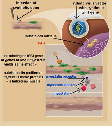
The
Performance Enhancing Drugs of the Future...an>
not steroids, but the
introduction of artificial
genes: Figure*
1. genes
for myosin type transcriptions
factors, that will activates genes
genes for long dormant myosin
isoforms of our ancient ancestors...
say an ancient type IIb
isoform
that's faster than any known
Type II isoform of today
2. or
IGF-I
(insulin-like growth factor)
IGF-I is a growth factor
structurally related to insulin and IGF-I is produced in
response to GH and then
induces subsequent cellular activities, particularly
on bone growth. IGF-I
has autocrine and paracrine activities, and like the
insulin receptor, it has intrinsic tyrosine kinase
activity. Owing to their structural similarities IGF-I
can bind to the insulin receptor.
next
Muscle Cell Growth includes:
1. satellite cell recruitment*, which proliferate & fuse with
muscle cell fibers
2.
pro-growth factors as IGF-I, which promotes satellite cell
proliferations
3.
growth inhibition factors, such as myostatin
Current research - H.L. Sweeney
at U. Penn
have used adeno-associated
virals (AAV) to infuse
IGF-I gene*
into recipient muscle cells
in normal mice:
experiments have overall size & growth rates
up 15% to
30%
in mice genetically engineered to
overproduce IGF-I: seen 20% to 30%
larger muscle mass
overproduction
also hastens muscle repair in mice with M.D.
injection of AAV-IFG-I into one leg of lab rats with an 8
week weight training program
= 2x increase in strength in treated leg
= longer period before gained strength is
lost
= sedentary rats showed 15% increased strength
Human trials for IFG-I injections to treat
myotonic (prolonged contraction) dystrophy
are set to begin next year...
next
to
be followed by athletic gene doping?
Myostatin...
is a muscle inhibitory
growth factor [blocks muscle growth],
myostatin
is also called
GDF-8 (growth differentiation
factor)
it promotes atrophy and slow muscle cell
growth,
may
function
antagonistically with IGF-I, which promotes muscle growth. |
described by A.C.McPerrron & Se-jin Lee at
Johns Hopkins in 1997
defective myostatin genes = considerably larger muscle
mass
Belgian Blue cattle* and the
Breed
&
its
cause
a
human case study* --> |
 reference
|
may be useful in muscle debilitating
diseases,
which include:
muscular dystrophy -
sarcopenia - age realted muscle loss
cachexia - aggressive muscle loss in cancer
& HIV patients
myoclonus - abnormal muscle contractions |
Wyeth
pharmaceuticals is at work on myostatin inhibitors
1st drugs
to date are antibodies to myostatin and
some clinical trials are set to begin in M.D. patients
|
| end
|
ATP
contraction cycle*
Training, Muscle
Fiber
Recruitment, & Performance & Marathoner pics
Muscle
Performance,
Training, & Fiber Recruitment
Disuse of a muscle, as in space travel
(weightlessness),
or
a couch potato can shrink
a muscle by 20% in 2
weeks.
Weight Training can increase
muscle mass to 150% of
normal size.
How do muscles get bigger and
better?
by making more muscle proteins...
nuclei of muscle control translation,
thus
one needs more nuclei, but muscle cell nuclei don't
divide.
New nuclei come from independent adjacent cells (Satellite stem
cells*).
when muscles under rigorous exercise they "tear", and the damaged
area
attracts satellite cells
into the tears, depositing more nuclei.
weight training thus leads to
heterotrophy of muscles......
more nuclei equals muscle
enlargement due to more protein synthesis.
next
Recruitment of Muscle Fibers (Slow
<--->
Fast) Is it Possible
?
has implications for spinal injury & athletics
1.
Cross innervation:
experimentally switch nerve innervations (slow to
fast)
animal experiments have lead to some conversions
2. Spinal
injury: a lack of nerve impulse &
muscle atrophy leads to a sharp
decrease of the slow myosin isoform (type I),
while the amount of the fast
isofrom increases (type II)
table*
Conclusion: neural input (electrical
stimulation) is necessary for the
proper
genetic expression of the Slow
- Type I isoform.
Electrical
stimulation can reintroduce the slow fiber into
paralyzed
muscles.
next
3. Weight
Training and Different
Myosin Types
sedentary people have higher
amounts of IIx
active people have more IIa fibers
heavy weight-load
repetitions.....
decreases Fast IIx
fibers and converts them to Fast
IIa fibers
nuclei stop expressing IIx
gene and express IIa
genes
after 1 month all IIx
--> IIa (muscle
also become more massive)
4. Tapering - can
we change amounts of IIx
fibers?
in experiments involving sedentary young
adults:
heavy resistance training (3 months) reduced IIx from 9% to 2%
but, a taper (rest for 3 months)
& IIx returned above
basline (9%)
to a level of 18%,
i.e., more fastest twitch
fibers. fig
5. Can we recruit slow
---> fast ? maybe...
but no good evidence to
date for slow to fast recruitments.
next
&
muscle
filament nomenclature
a protein called PPAR-delta,
discovered by Ron Evans of Salk Institute
regulates other genes involved in fat metabolism.
High activity of PPAR-d burns more fat,
results in leaner, more fit individuals.
recent experiments (PLoS
- Oct 2004)
revealed that mice genetically
modified to produce more
PPAR-d
- had 2x more slow twitch (I) muscle compared to
litter-mates. [fig]*
- PPAR-d mice could run 1,800m (2x normals) before
reaching exhaustion.
these changes are similar to those induced by
sustained training & exercise
long lasting vigorous exercise produces a higher ratio
od slow twitch (I) muscle.
a new drug (GW501516) activates PPAR-d
directly leading to similar changes
- could help obese and heart disease patients who
can't exercise.
GlaxoSmithKline is currently testing this drug in
obese, diabetics
end
7. Why muscles deteriorate
with age
|
|
xxx
x
|
 |
|
the end.
el
fin,
de ende,
il finito
elnihaya,
telos,
finis,
au
revoir
farvel a hui hou.
|
| best in long
slow sustained contractions |
best
in rapid (short) contractions |
| not easily
fatigued |
easily fatigued |
| more capillary
beds, greater VO2 max |
less capillary beds |
| smaller in size |
larger in size |
| lower
glycogen content |
higher glycogen content |
| poor
anaerobic glycolysis |
poor
but some aerobic capacity |
| higher fat content |
lower fat content |
| more
mitochondria - Beta Oxidation high |
fewer
mitochondria- Beta Oxidation low |
| poorly formed sarcoplasmic reticulum |
well formed sacroplasmic reticulum |
| slower release of Ca = slower contractions |
quick release of Ca = rapid contractions |
| tropinin has lower affinity for Ca
|
troponin - higher affinity for Ca |
atp cycle
https://www.youtube.com/watch?v=gJ309LfHQ3M
|
