NEUROPHYSIOLOGY...
material from Chapter 48 & 49.
Cell Communication... at the
molecular levell --> SIgnal
Transduction Mechanism.
via Electrical Properties of Nerve cells
(neurons)...
the electrophysiology of neurons
lies in their... Membrane
Physiology
there is a great
diversity* of
Nervous Systems [mouse
gut & mouse retinal
ganglia]
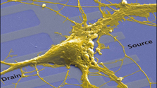
a neurophysiology model organism is
: Squid*Giant Axon
[Doryteuthis pealeii]
   |
NERVE CELL FUNCTION
- Reception-Transduction-Response*
is akin to SIGNAL TRANSDUCTION
: reception -
transduction - response
and a HOMEOSTATIC
REGULATOR
: receptor
- controller - effector
1. sense organs
via
Peripheral
NS gathers sensory
input --> signals info in
2. integration (transduces)
information (within
CNS - brain & spinal cord)
 3. responds via motor output (effector organs
& PNS
- muscles :
)
3. responds via motor output (effector organs
& PNS
- muscles :
) |
| |
a
common pathway in a Nervous System*
Reflex Arc - hard wired, unconscious rapid
response to external stimulus
involving spinal nerves
& effector cell electrical impulses
narrated
explanation of spinal reflex arc.*
knee-jerk reflex*
or
another
spinal reflex
To study the actions and functions of nerves and muscles
a good model
experimental system is the...
neuro muscular*junction

| |
|
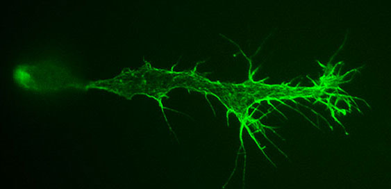
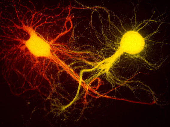
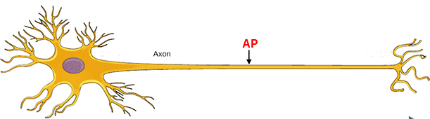
STRUCTURE of an
individual NERVE CELL [a NEURON*]
the term neuron was coined
by the German anatomist Heinrich von
Walder-Hartz
in 1891 to
describe an individual cell in the brain. |
 |
Cell
Body*
- is main part of neuron cell with cytoplasm & organelles
Dendrites*
- short cylindrical outgrowths of Cell
Body
carry signals (electrical impulses) into cell body
Axon* (pic)
- long outgrowth of cell body - carry signals
to next neuron
&
Schwann
cell - cells surrounding
peripheral neuron axons in vertebrates -
produce myelin (sheath)
membrane (mylein origins?*)
protein + lipid-like membrane insulating
material surrounding axon
Nodes
of Ranvier* - space between
successive Schwann cells along axon...
node area is a non-myelinated area that
regulates the
speed of
conduction -
w/o myelin speed is less (5 m/sec)
w/myelin (100 m/sec or 200 mi/hr)
Multiple
Sclerosis - degenerative
disease of myelin sheaths
Synaptic
Knob* - enlarged ends
of neuron holds neurotransmitters
in synaptic vesicles
Glia cells - (astrocytes) provide
support, function as blood-brain barrier,
etc...

|
What is the
informational content of neurons???
it's in the
ELECTRICAL PROPERTIES of neuronal cells...
What is the the
electrical charge across a cell's membrane...
what is
a RESTING & ACTION potentials
RESTING POTENTIAL
- is a characteristic ELECTRIC CHARGE
exhibited by cells at rest...
most often inside NEGATIVE
(-) in animal cells
which is due to the existing
distribution of ions across the cell
membrane.
electrical potential - (in electrical
terms) is amount of
electrical charge at one point
in an electric circuit compared to some other
point in the same
circuit, often measured with a volt-meter (multi-meters*)
How to measure
membrane potentials* in cells -
microelectrode*
recording
equipment*

|
inside vs.
outside of cells
|
| SGA |
- |
65 to - 70
mVi |
| Frog muscle fibers |
- |
90 mVi |
| Nitella
(algae) |
- |
150 mVi |
| Valonia (algae) |
+ |
15 mVi |
| |
|
|
Causes
of Resting Potential... what is cause of
inside -65 to -70 mVi for SGA ?
homeostatic distribution of ions
in neurons & surroundings*
all of which result in the inside of cell
being negative (-65mV) But HOW ?
how are these ions distributed across a SGA - passively or actively?
| |
ENa
= +62mVi
150/15 = lg 10
= values* |
|
| Nernst Eq.* Emv=
+/-62 lg10[Co]/[Ci] |
EK =
-90mVi
5/140 = lg 0.035
|
|
| |
ECl
= -67mVi
120/10 = lg 12
|
|
Ion transport through channel
proteins establishes the Resting Membrane Potential
include:
1.
active transport of Na & K* high Na outside [3] & high K inside [2] via NaK-ATPase
2. differential
permeability* diffusion of K (faster out)
& Na
(slower in) = inside (-)
3. lots of protein anions (-)
at cell pH, thus inside more (-) then outside
4. normal
diffusion
of Cl-
into the cell
ACTION
POTENTIAL... change resting potential
charge due to passive ion transport
9e: c4
-
name given to changes in
electrical charges occurring during
stimulation of a nerve cell,
- transient
change in the RP voltage at one point along membrane of nerve
cells...
- change in charge is
self-propagating along an axon
- usually visualized
graphically from an oscilloscope* recording [ graph*of AP*]
PROPERTIES of an AP...
... requires a living cell,
i.e., requires O2 for metabolism
is eliminated by metabolic poisons, including
cyanide
... requires Na/K pump
and ion
channels for
Na & K (fig*) to produce ion
distributions
... is measured at local point using microelectrodes* impaled in cells or a
nerve chamber*
... has a threshold
- a minimal amount of
stimulus is needed to open channels &
"fire" an AP
... is an "all-or-none"
phenomena, either yes or no, but is
always of same magnitude
... is very rapid - time course = 2-3 msec to
elicit change in polarity

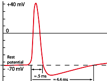
EVENTS DURING an AP oscilloscope trace*
graph of AP
Voltage changes
the permeability of axonal membranes and
elicit an Action Potential.
depolarization* - cell goes
from inside negative
(-) to inside
positive (+)
Na channel opens - Na
diffusively floods in --> -70mV toward +62mV
repolarization* -
Na channels close & K channels open outward
[returns to inside (-)]
K
follows its diffusive gradient & K
diffuses out of cell (returns -)
hyperpolarization
-
"undershoot or overshoot"
of resting potential (-75mv)
refractory
period -
time before another AP can "fire" again (about
2-3 mSec)
overall mechanism*** &
AP
animation (Pearson)*
Current
changes during an AP
CONDUCTION of an AP
along an axon
local spreading of electric charge depolarizes adjacent
membranes
changes the membrane permeability of adjacent
non-myleinated region
leading to continuous propagation in an autocatalytic - "domino
effect"...
figure*
AP Conduction is continuous in
non-mylinated
axons
=
2-3 m/sec
Saltatory Conduction* in mylinated axons
[node to node] = 100 m/sec
Synaptic Transmission... synapse animation*
.
synapse - functional
space connecting two
neurons allowing transmission of
AP's between cells: may be electrical or chemical
synaptic cleft -
[20 to 30 nm across] open space between neurons
across which a chemical neurotransmitter may
diffuse
synaptic knob
- site of vesicles
holding neurotransmitter at end of axon
synaptic vesicle - holds
neurotransmitters (ex: acetylcholine in
neuromuscular junctions)
action potentials
trigger release of neurotransmitters*
pre-synaptic side - releases
neurotransmitter
post-synaptic side - has
a receptor which
binds transmitter and ...
ion channels open -
leads to change of potential charge
on the post-synaptic membrane ----> new AP
removal
of stimulus - an enzyme
destroys neurotransmitter: ex. "ACH-ase"
Post-synaptic
membrane potential responses...
depends upon post-synaptic receptor type and
how it responds.
EPSP -
excitatory post-synaptic
potential [goes positive from RP
[-65mVi] to -55mVi]
excitatory PSM neurons -->
open Na channels
--> inside +
--if threshold--> AP
figure*
IPSP -
inhibitory post-synaptic potential
[goes negative from RP to
-80mVi]
inhibitory PSM neurons --> opens Cl channels - Cl-in
-> more - --> no
AP
--> opens K
channels - K-out ->
more - --> no
AP
AP - all or none 120mv
depolarization/repolarization (from -65 to +55 mVi)
Neurotransmitters... some stimulators
and drugs.
| neuro-muscular junction |
acetylcholine... muscle
contractions
[cholinergic neurons = Na+ influx] |
biogenic amines
(CNS & PNS) |
epinephrine &
nor-epinepherine - [catecholamines]...
increase heart rate
serotonin &
dopamine
- affect
mood, attention, & learning
depression = often
from reduced epinephrine/norepinephrine
levels
or an imbalance of serotonin.
|
| amino acids |
ASP & GLU -
excitatory (CNS)
Chinese Restaurant
Syndrome +
MSG
GLY
& GABA
-
inhibitory (Cl-) |
peptides
(small proteins) |
endorphins
- their role [discovery] is to
decrease perception of pain
substance-P
-
excitatory transmitter - signaling pain |
Stimulants/Depressants - are chemicals that effect
the activities of neurons...
|
cocaine
- blocks re-uptake
of* dopamine by synaptic
vesicles --> continual stimulation
of
reward-motivated behavior
animation
of cocaine action
|
flea collars -
fipronil
- blocks GABA-gated*
Cl channels (normally inhibitory),
thus preventing hyperpolarization = results in
excessive neural excitation,
a hyper-excitation of
CNS of the fleas and death.
|
strychnine
(a poison) - antagonist of
inhibitory GLY &
ACH receptors in spinal cord,
resulting in increase
muscular convulsion & asphyxia.
|
Sarin
(a nerve agent) blocks
action of
acetylcholinesterase,
thus
overloading body's
nervous system in
toto, ultimately paralyzing the
diaphragm = suffocation.
|
 Key Concepts*
synaptic
cleft - the Glut-Tang Clan video Key Concepts*
synaptic
cleft - the Glut-Tang Clan video |
END
copyright c2024
Charles Mallery,
Biology 150, Department of
Biology, U. of Miami, Coral
Gables, FL 33124
|
copyright c2024
Charles Mallery, Biology
150, Department of Biology, U. of
Miami, Coral Gables, FL 33124
SKIP the MATERIAL
BELOW
NRC-Summary
STRUCTURAL PARTS of Nervous
System -
central
nervous system - brain
and spinal cord...
(neural
stem cell = origin of brain)
peripheral nervous system - outside the CNS-
carries signals in/out
of CNS
PNS =
sensory (affernet
- in)
and motor (efferent - out)
neurons:
- somatic nervous system - carries signal to skeletal
muscle - under conscious
control
- autonomic
nervous system - regulate homeostatic internal systems - involuntary control
2 complimentary
Parasympathetic (cranial & cholenergic)
- calming: HR-, energy
storage
systems: Sympathetic (spinal & noradrenaline) -
energy & arousal: HR+, glycogen--> glu
FUNCTIONAL TERMINOLOGY of
Neurons -
Nerve
- bundle of individual
neuron cells wrapped in connective tissue
Ganglia - cluster of cell bodies of
individual neurons
Sensory neurons... (afferent
neurons) -
external stimuli from receptors toward CNS
Interneurons... integrate & relay sensory
input to motor neuron
Motor
Neurons... (efferent
neurons) -
convert signals to effector
cells = response

Neurotransmitters
& Other stimulators and
drugs...
Some
common Neurotransmitters...
Table of transmitters*
Harvey Project
Dr. King's site
Prozac
and Paxil
(antidepressants)...
animation
blocks reabsorption of serotonin from synaptic cleft
& boost synthesis of serotonin, epinepherine,
& norepinepherine
Parkinson's = lack of dopamine schizophrenia = too
much dopamine
LSD/mescaline - psycho-active drugs
function by
binding to serotonin/dopamine brain cell receptors
facilitates
juvenile brain plasticity
Campbell reads:
10e: c48 (pg 1061-1077) c49 (pg 1079-1083)
11e: c48 (pg
1065-1079) c49 (pg 1083-1088)
|

![Summary of SGA [ions] & resting potential](http://henge.bio.miami.edu/mallery/lec/150/neuro/48x5.jpg)